PET (Positron Emission Tomography)
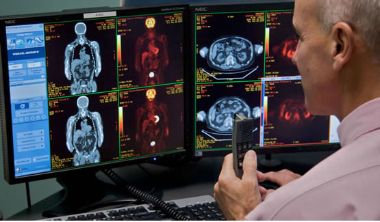
Accredited by the American College of RadiologyPET/CT simultaneously images and combines the results of two state-of-the-art scanner technologies into a single exam: the highly sensitive PET scan has the ability to detect changes in cell function and provides a detailed picture to reveal the size and shape of abnormal growths.
PET/CT is one of the most powerful tools that allows your physician to diagnosis cancer and other diseases, determine the best treatment plan and monitor your progress. It picks up actively growing cells, and the CT scan provides a detailed picture of the inside of your body to reveal the size and shape of abnormal growths.
At this time, PET/CT is one of the most powerful tools in cancer diagnosis and treatment planning. It has the highest sensitivity and specificity of any tumor detection study available. In addition, PET/CT can provide important information about some neurological conditions, such as Alzheimer's Disease as well.
PET Exam Prep
- Patients having a PET scan of the breast, lung and/or head must be NPO (fasting) for 4.5 hours prior to exam.
- Patients having any other PET scan are to be NPO (fasting) for 6 hours prior to exam.
- Diabetic patients are to call for special instructions: 772-562-0163 ext. 129.
- Patients will be called the day before your appointment to review specific instructions regarding your prep.
- Please note you will be given instructions on specific food groups you must include in your diet as well as ones to avoid.